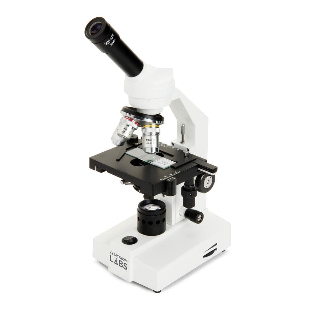
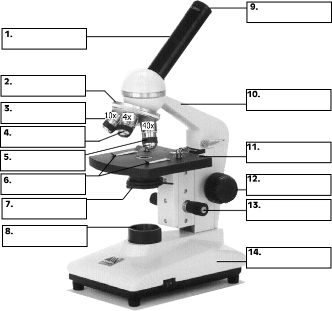
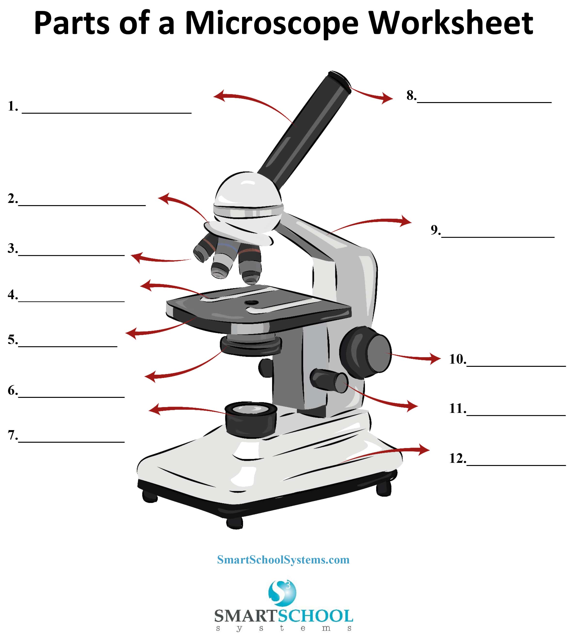
39 compound microscope diagram unlabeled
Join LiveJournal Password requirements: 6 to 30 characters long; ASCII characters only (characters found on a standard US keyboard); must contain at least 4 different symbols; Lifestyle | Daily Life | News | The Sydney Morning Herald The latest Lifestyle | Daily Life news, tips, opinion and advice from The Sydney Morning Herald covering life and relationships, beauty, fashion, health & wellbeing
Parts of the Microscope with Labeling (also Free Printouts) 5. Knobs (fine and coarse) By adjusting the knob, you can adjust the focus of the microscope. The majority of the microscope models today have the knobs mounted on the same part of the device. Image 5: The circled parts of the microscope are the fine and coarse adjustment knobs. Picture Source: bp.blogspot.com.

Compound microscope diagram unlabeled
Microscope Diagram Teaching Resources | Teachers Pay Teachers Teaching to the Middle. 4.8. (24) $1.50. PDF. This passage briefly describes microscopes and their parts (900-1000 Lexile). 14 questions (matching and multiple choice) assess students' understanding. Students label a diagram of 6 parts of a microscope. I've included a color and BW version, as well as a key. Compound Microscope Parts - Labeled Diagram and their Functions The term "compound" refers to the microscope having more than one lens. Basically, compound microscopes generate magnified images through an aligned pair of the objective lens and the ocular lens. In contrast, "simple microscopes" have only one convex lens and function more like glass magnifiers. Elodea Leaf Cell Diagram Nov 04, 2019 · Under the Microscope Have students determine the field diameter of the compound microscope objectives. This Elodea leaf cell exemplifies a typical plant cell. It has a nucleus, and a stiff cell wall which gives the cell its box-like shape. The numerous green chloroplasts allow the cell to . Minecraft Circle Diagram. Standing Rigging Diagram.
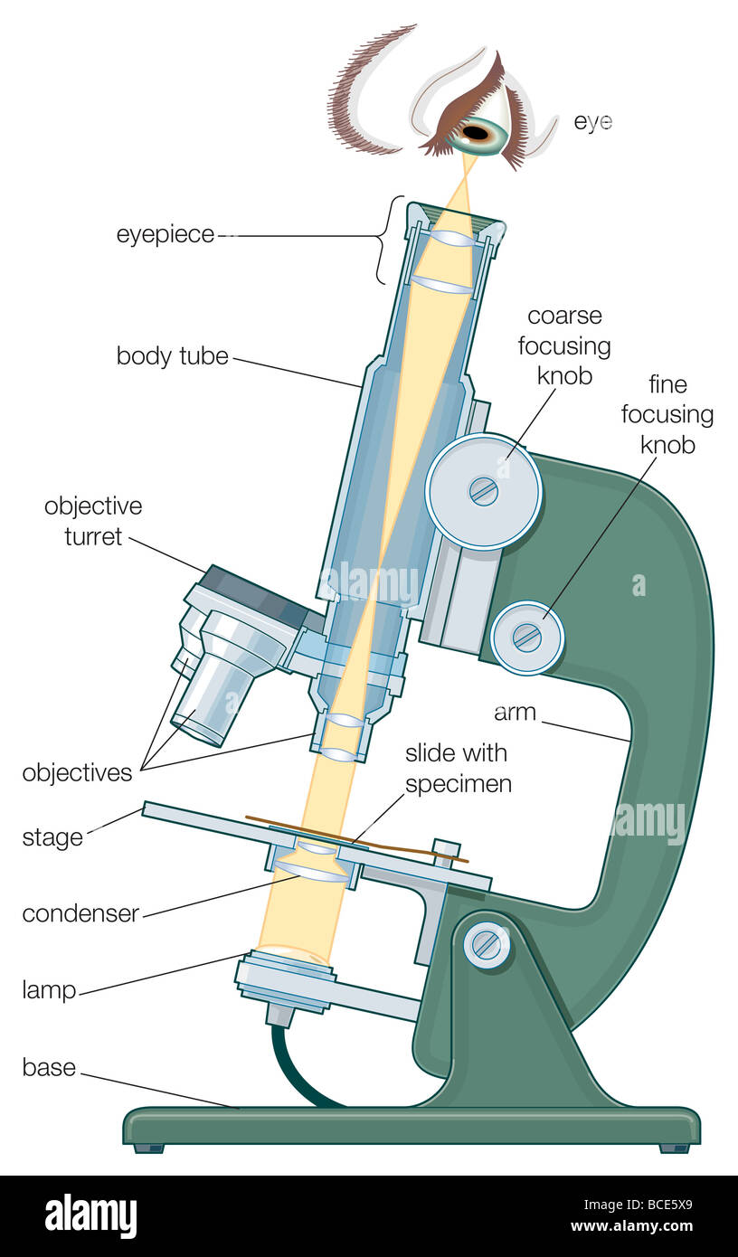
Compound microscope diagram unlabeled. Microscope Types (with labeled diagrams) and Functions A compound microscope: Is used to view samples that are not visible to the naked eye Uses two types of lenses - Objective and ocular lenses Has a higher level of magnification - Typically up to 2000x Is used in hospitals and forensic labs by scientists, biologists and researchers to study micro organisms Compound microscope labeled diagram Parts of a microscope with functions and labeled diagram - Microbe Notes Figure: Diagram of parts of a microscope There are three structural parts of the microscope i.e. head, base, and arm. Head - This is also known as the body. It carries the optical parts in the upper part of the microscope. Base - It acts as microscopes support. It also carries microscopic illuminators. Label the microscope — Science Learning Hub All microscopes share features in common. In this interactive, you can label the different parts of a microscope. Use this with the Microscope parts activity to help students identify and label the main parts of a microscope and then describe their functions. Drag and drop the text labels onto the microscope diagram. Compound Microscope- Definition, Labeled Diagram, Principle, Parts, Uses Alternatively, the magnification of the compound microscope is given by: m = D/ fo * L/fe where, D = Least distance of distinct vision (25 cm) L = Length of the microscope tube fo = Focal length of the objective lens fe = Focal length of the eye-piece lens Parts of a Compound Microscope Eyepiece And Body Tube.
A phosphate starvation response-centered network regulates ... (A) The schematic diagram shows the position of P1BS in OsRAM1, OsWRI5A, OsPT11 and OsAMT3;1 promoters. (B) EMSA assay showing specific binding of OsPHR2 to the P1BS element. The unlabeled mP1BS (GNAGAGNC) containing DNA fragment (10-fold, 50-fold, 100-fold) was used as a competitor. Compound Microscope Labeled Diagram | Quizlet Compound Microscope Labeled + − Flashcards Learn Test Match Created by meganplocher734 Terms in this set (14) Eyepiece/Ocular lens Contains the ocular lens Body tube A hollow cylinder that holds the eyepiece. Arm Part that supports the microscope. Stage Supports the slide or specimen Coarse adjustment Knob Compound Microscope Diagram Blank - 16 images - types of microscopes ... Thursday, May 12, 2022 microscope diagram labeled unlabeled and blank parts of a microscope Compound Microscope Diagram Blank. Here are a number of highest rated Compound Microscope Diagram Blank pictures on internet. We identified it from reliable source. Its submitted by presidency in the best field. Compound Microscope: Definition, Diagram, Parts, Uses, Working ... - BYJUS What is a Compound Microscope? A compound microscope is defined as A microscope with a high resolution and uses two sets of lenses providing a 2-dimensional image of the sample. The term compound refers to the usage of more than one lens in the microscope. Also, the compound microscope is one of the types of optical microscopes.
Mapping Genomes - Genomes - NCBI Bookshelf Draw a diagram showing how a double-stranded cDNA is synthesized. 15. Define the term ‘mapping reagent’ and explain how a panel of radiation hybrids is used as a mapping reagent. 16. Explain how a clone library is used as a mapping reagent. 17. Draw a diagram to show how a sample of a single human chromosome can be obtained by flow cytometry. Compound Microscope - Diagram (Parts labelled), Principle and Uses See: Labeled Diagram showing differences between compound and simple microscope parts Structural Components The three structural components include 1. Head This is the upper part of the microscope that houses the optical parts 2. Arm This part connects the head with the base and provides stability to the microscope. Microscope Diagram Labeled, Unlabeled and Blank - Pinterest When students map a microscope, they assign colors and/or patterns to each part they need to identify. They fill in the key and the corresponding part. This file includes two types of microscope maps: 1. a microscope with the twelve parts listed along the side 2. a microscope with arrows that allows the student to… J Jackie Schoen Teaching Things PDF AN INTRODUCTION TO THE COMPOUND MICROSCOPE - Rowan University AN INTRODUCTION TO THE COMPOUND MICROSCOPE OBJECTIVE: In this lab you will learn the basic skills needed to stain and mount wet slides. You will also learn about magnification, resolution and the parts of the compound microscope. INTRODUCTION: The light microscope can extend our ability to see detail by 1000 times, so that we can
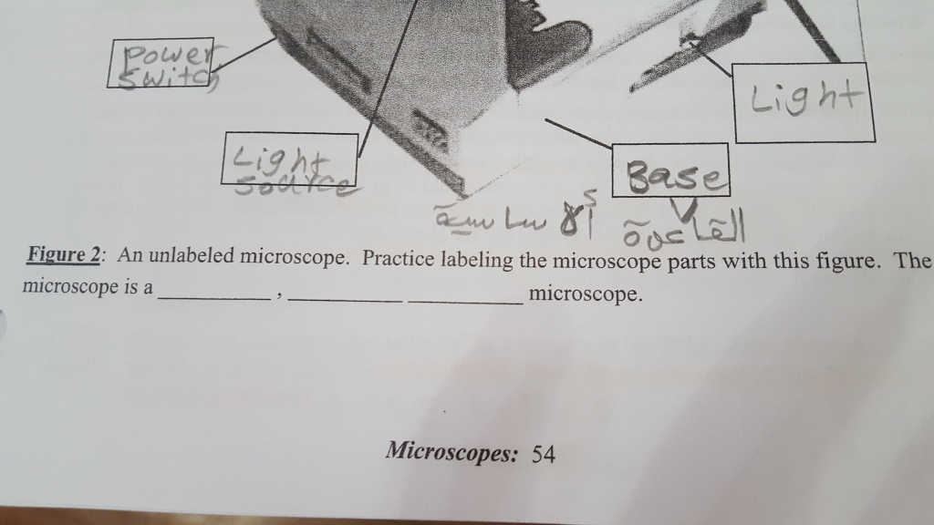
A Study of the Microscope and its Functions With a Labeled Diagram ... Here, unlabeled microscope diagrams have been provided for your perusal, which will help you practice and test your understanding of the instrument. Types of Microscopes Depending on the source of illumination, microscopes can be divided into two categories. They are:
The compound microscope - how to draw ray diagrams - YouTube An animated presentation showing you how to draw ray diagrams (using simple lens rules) for a compound microscope. This shows how to determine the position a...
Binocular Microscope Anatomy - Parts and Functions with a Labeled Diagram First, see the body and arm of the light compound microscope. The body tube is a cylindrical-like structure that connects the ocular lens to the objective lenses. Again, the arm of the microscope connects the body tube to the microscope's base. You will see the coarse and fine adjustment in the arm of the microscope.
Diagram of a Compound Microscope - Biology Discussion A bright-field or compound microscope is primarily used to enlarge or magnify the image of the object that is being viewed, which can not otherwise be seen by the naked eye. Magnification may be defined as the degree of enlargement of the image of an object provided by the microscope.
Looking at the Structure of Cells in the Microscope ... A light microscope. (A) Diagram showing the light path in a compound microscope. Light is focused on the specimen by lenses in the condensor. A combination of objective lenses and eyepiece lenses are arranged to focus an image of the illuminated specimen
Draw a neat labelled diagram of a compound microscope and ... - Sarthaks Using sign convention, we find that O'I 1 = + v 0 and O'O = -u where v 0 is the image distance due to the objective and u is the object distance for the objective or the compound microscope. I 1 G 1 is negative and OJ is positive. To find me : The eyepiece behaves like a simple microscope. So : the magnifying power of the eye piece. ∴ m e ...
How to draw Compound Microscope Diagram - Final Image at D Learn to draw Compound Microscope Diagram (Final Image at least distance of distinct vision "D") within 3 mins.Background Score Credit: Energy from ...
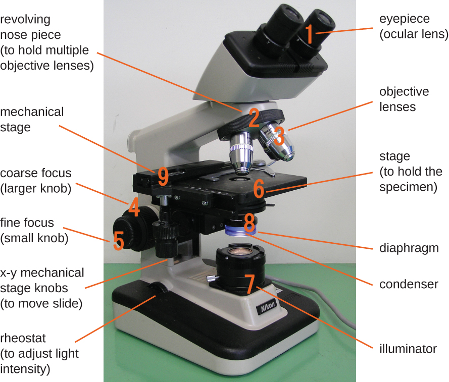
Parts of a Compound Microscope and Their Functions - NotesHippo Compound microscope mechanical parts (Microscope Diagram: 2) include base or foot, pillar, arm, inclination joint, stage, clips, diaphragm, body tube, nose piece, coarse adjustment knob and fine adjustment knob. Base: It's the horseshoe-shaped base structure of microscope. All of the other components of the compound microscope are supported ...
Compound Microscope - Types, Parts, Diagram, Functions and Uses It comes with a wide body and base. Its distinct parts include a condenser, illumination, focus lock, mechanical stage, and a revolving nosepiece which can hold up to five objectives. It usually has a binocular head, which makes long-term observation easy. Image 22: An example of a research compound microscope.
Compound Microscope Drawing : Microscope Diagram Labeled Unlabeled And ... Print a microscope diagram, microscope worksheet, or practice microscope quiz in order to learn all. 10 1 3 the compound microscope. Compound microscope drawing vine drawing, body drawing, woman drawing,. The type of microscope in our drawing guide is known as a compound microscope, an optical microscope, or a light microscope.
Draw a labelled ray diagram of a compound microscope and ... - Vedantu In a compound microscope the focal length of the objective is 0.5 cm and the focal length of eyepiece is 5 cm. The real image of the object is formed at a distance of 15.5 cm from the objective. If the final image is formed at a distance of 25 cm from the eyepiece, what is the magnifying power of the microscope? A. 180 B. 0.2 C. 20 D. 150
Junqueira's Basic Histology Text and Atlas, 14th Edition There is shortage of references in higher teaching institutions especially in newly opened institutions engaged in training of various Veterinary professionals in the country.
Labelled Diagram of Compound Microscope The below mentioned article provides a labelled diagram of compound microscope. Part # 1. The Stand: The stand is made up of a heavy foot which carries a curved inclinable limb or arm bearing the body tube. The foot is generally horse shoe-shaped structure (Fig. 2) which rests on table top or any other surface on which the microscope in kept.
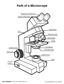
Microscope Diagram Labeled, Unlabeled and Blank | Parts of a Microscope ... Microscope Diagram Labeled, Unlabeled and Blank | Parts of a Microscope Print a microscope diagram, microscope worksheet, or practice microscope quiz in order to learn all the parts of a microscope. Tim's Printables 39k followers More information Microscope Diagram Find this Pin and more on Science Printables by Tim's Printables.
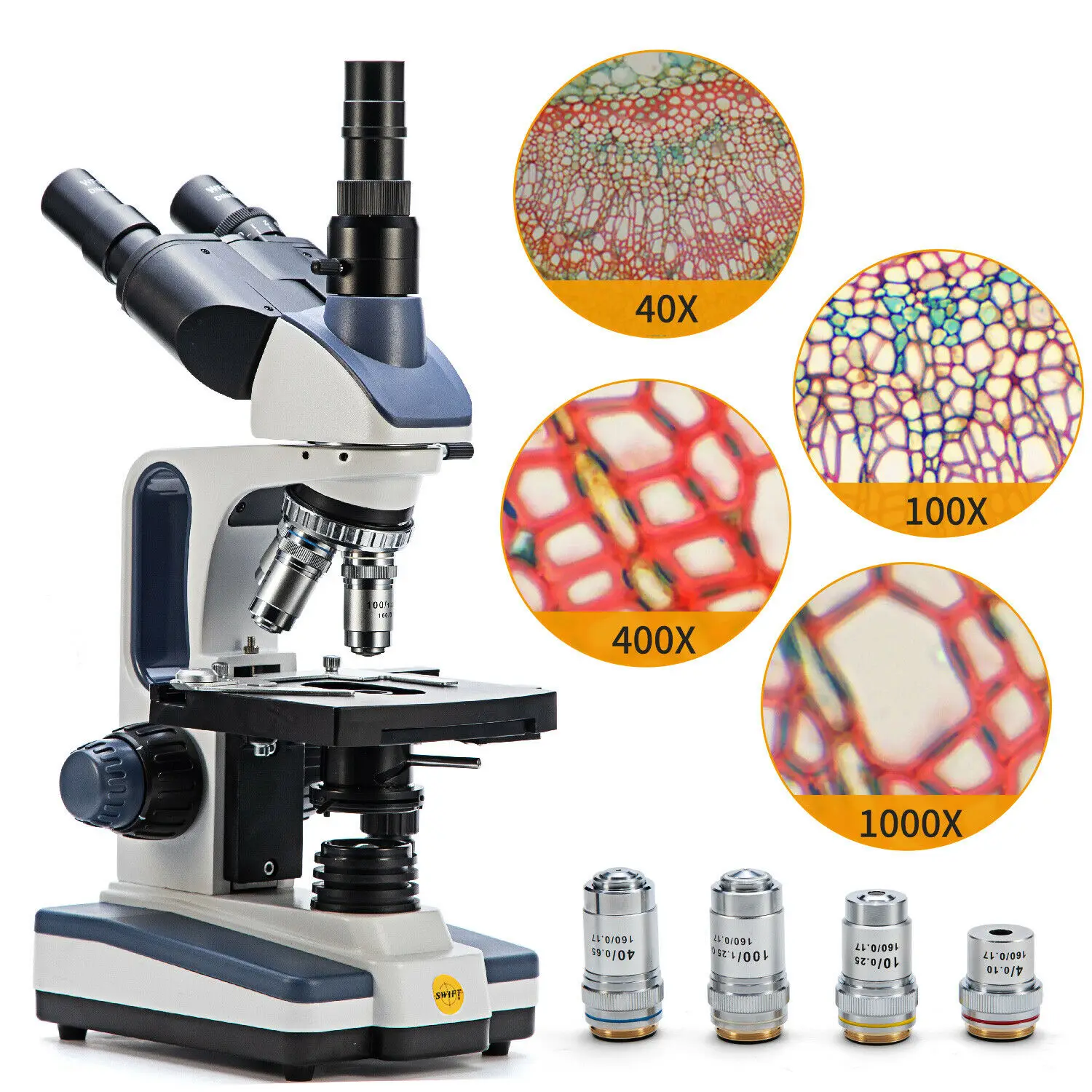
Microscope Parts and Functions First, the purpose of a microscope is to magnify a small object or to magnify the fine details of a larger object in order to examine minute specimens that cannot be seen by the naked eye. Here are the important compound microscope parts... Eyepiece: The lens the viewer looks through to see the specimen.
16 Parts of a Compound Microscope: Diagrams and Video Body of the Microscope In compound microscopes with two eye pieces there are prisms contained in the body that will also split the beam of light to enable you to view the image through both eye pieces. 2. Arm The arm of the microscope is another structural piece. The arm connects the base of the microscope to the head/body of the microscope.
Compound Microscope Parts, Functions, and Labeled Diagram The total magnification of a compound microscope is calculated by multiplying the objective lens magnification by the eyepiece magnification level. So, a compound microscope with a 10x eyepiece magnification looking through the 40x objective lens has a total magnification of 400x (10 x 40).
labeling a microscope worksheet A Study Of The Microscope And Its Functions With A Labeled Diagram . mikroskop bagian fungsinya microscope cahaya jenis lensa lengkap fungsi makalah binokuler okuler jelaskan elektron perbedaan labeling. Microscope Diagram Labeled, Unlabeled And Blank | Parts Of A Microscope . microscope compound ...
Elodea Leaf Cell Diagram Nov 04, 2019 · Under the Microscope Have students determine the field diameter of the compound microscope objectives. This Elodea leaf cell exemplifies a typical plant cell. It has a nucleus, and a stiff cell wall which gives the cell its box-like shape. The numerous green chloroplasts allow the cell to . Minecraft Circle Diagram. Standing Rigging Diagram.
Compound Microscope Parts - Labeled Diagram and their Functions The term "compound" refers to the microscope having more than one lens. Basically, compound microscopes generate magnified images through an aligned pair of the objective lens and the ocular lens. In contrast, "simple microscopes" have only one convex lens and function more like glass magnifiers.
Microscope Diagram Teaching Resources | Teachers Pay Teachers Teaching to the Middle. 4.8. (24) $1.50. PDF. This passage briefly describes microscopes and their parts (900-1000 Lexile). 14 questions (matching and multiple choice) assess students' understanding. Students label a diagram of 6 parts of a microscope. I've included a color and BW version, as well as a key.








Post a Comment for "39 compound microscope diagram unlabeled"