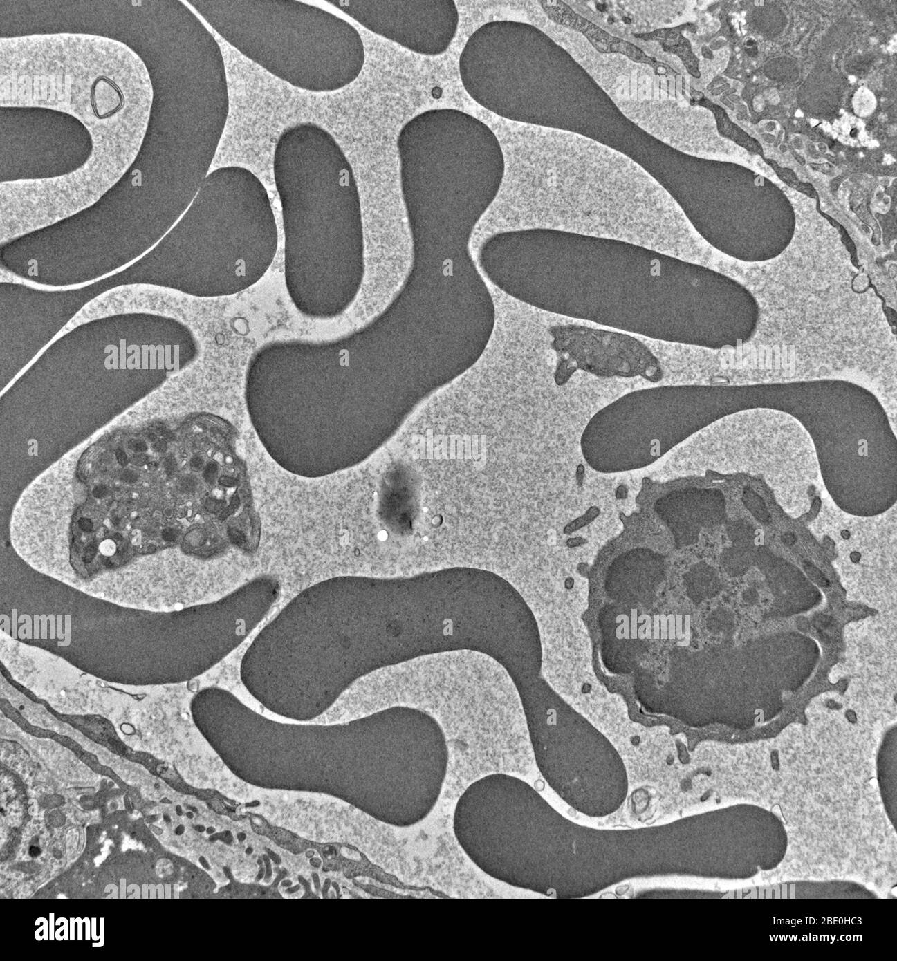
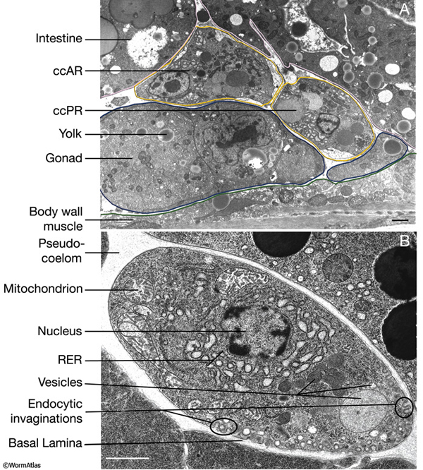
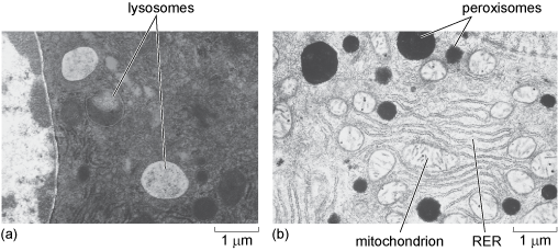
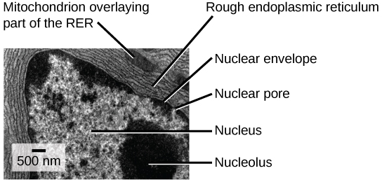
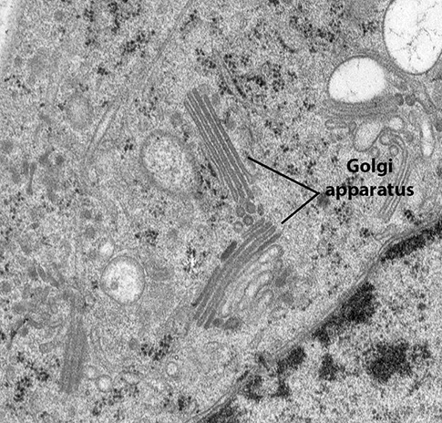
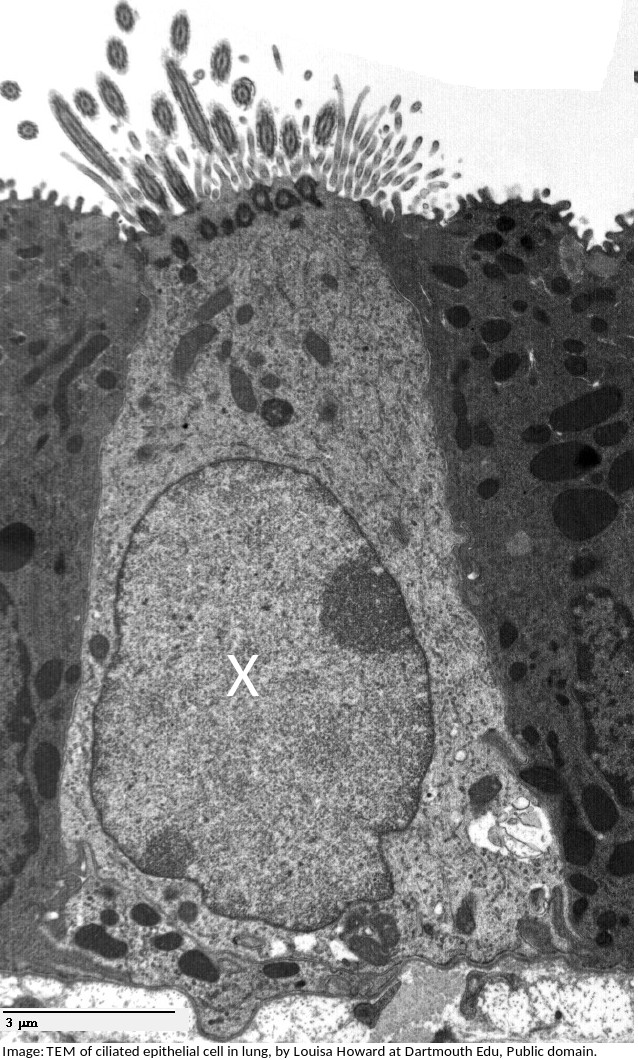
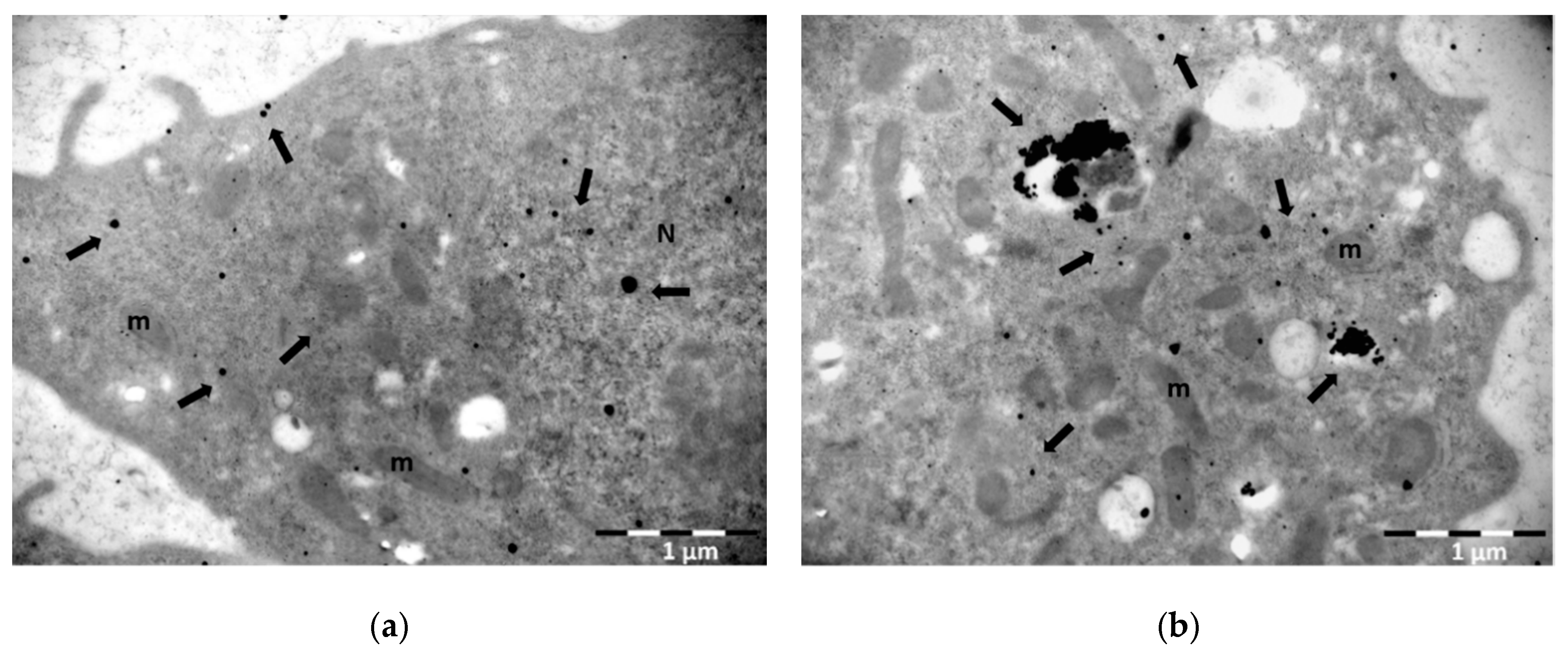
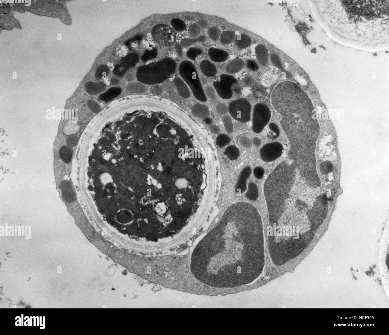
42 label the transmission electron micrograph of the cell.
Label This Transmission Electron Micrograph / Microscopy Innovations ... Label the transmission electron micrograph of the cell. Interpretation of electron micrographs to identify organelles and deduce the functions of specialized cells. In electron microscopy, however, true genetic encoded multilabeling. Label the transmission electron micrograph of the nucleus. animal cell under electron microscope labelled - Be A Terrific Memoir ... It is an electron micrograph of cells largest and most important organelle the mitochondria and is characterized by the following features Fig. A typical animal cell is 1020 μm in diameter which is about one-fifth the size of the smallest particle visible to the naked eye. When observing onion cells there is the Cell Surface.
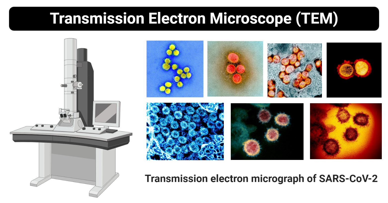
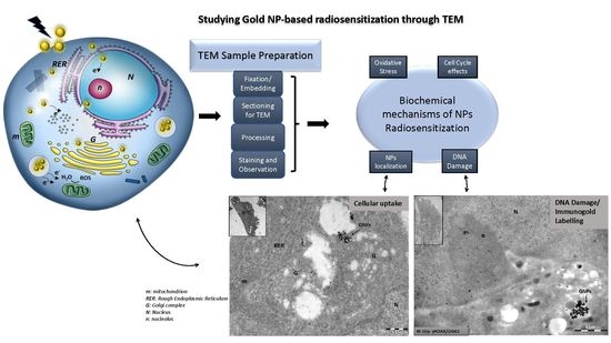
Transmission Electron Microscope (With Diagram) - Biology Discussion Finally, the electrons are focused by an electromagnetic projector lens (instead of an ocular lens as in a light microscope) on a screen or photographic plate. The final image in a TEM is known as transmission electron micrograph. The salts of some heavy metals, e.g., lead; osmium, tungsten and uranium are often used for staining.

Label the transmission electron micrograph of the cell.
Labeled Plant Cell Under Electron Microscope : The Cell Form 1 Biology ... Under a microscope, epithelial cells are readily distinguished by the following features: The electron microscope can achieve a resolution of up to 100 picometers, allowing eukaryotic cells, prokaryotic cells, viruses, ribosomes, and even single atoms to be visualized (note the in order for dna to be clearly visualized under an electron microscope, it must be labeled with heavy atoms. #6 Summary of Cell structure | Biology Notes for A level - Blogger 9 The electron micrograph shows part of a secretory cell from the pancreas. The secretory vesicles are Golgi vesicles and appear as dark round structures. The magnification is x 8 000. a Copy and complete the table. Use a ruler to help you find the actual sizes of the structures. Give your answers in micro metres. Electron Microscopic Study of Cell and Organelles - GK SCIENTIST Some of the membranes of endoplasmic reticulum open out in the cell membrane and the others in the nuclear membrane. Golgi apparatus in cytoplasm appears, under an electron microscope, as a pile of 2-layered flattened sacs called cisternae or saccules, lying one within the other.The sacs have flattened or swollen ends near which lie the vesicles of varying size and shape.
Label the transmission electron micrograph of the cell.. Transmission electron micrograph of epidermal Langerhans cells ... Transmission electron micrograph of epidermal Langerhans cells. (x89,000.) Colocalization of BL6-AU 15 and of BL2-AU 5 (arrows) in the same Birbeck granules. (A) The double labeling of the central... Transmission Electron Micrograph of transfected HL-1 cells labeled for ... Transmission Electron Micrograph of transfected HL-1 cells labeled for TMEM43 with immunogold. A and B. Single immunogold labeling experiments used 15 nm gold particles to label GFP. A.... Label the transmission electron micrograph of the cell. 0 Nucleus ... Label the transmission electron micrograph of the cell. 0 Nucleus rences Mitochondrion Heterochromatin Peroxisome Vesicle ULAR bumit Click and drag each label into the correct category to indicate whether it pertains to the cytoplasm or the plasma membrane. ICF Contacts the ECF Made of proteins and... › cell › fulltextBacPROTACs mediate targeted protein degradation in ... - Cell Jun 03, 2022 · As seen for pArg and cyclomarin head groups, various molecules that bind to the substrate receptor of the ClpCP protease can be incorporated into a functional degrader. Using cell permeable BacPROTACs, we furthermore demonstrate that recruitment of model proteins to ClpCP leads to selective protein degradation in bacterial cells.
en.wikipedia.org › wiki › Fluorescence_microscopeFluorescence microscope - Wikipedia Fluorescence micrograph gallery A z-projection of an osteosarcoma cell, stained with phalloidin to visualise actin filaments. The image was taken on a confocal microscope, and the subsequent deconvolution was done using an experimentally derived point spread function. Labeling the Cell Flashcards | Quizlet Label the transmission electron micrograph of the nucleus. membrane bound organelles golgi apparatus, mitochondrion, lysosome, peroxisome, rough endoplasmic reticulum nonmembrane bound organelles ribosomes, centrosome, proteasomes cytoskeleton includes microfilaments, intermediate filaments, microtubules Identify the highlighted structures › news › national-newsCDC releases new guidance on monkeypox and schools 2 days ago · Read More FILE – This image provided by the National Institute of Allergy and Infectious Diseases (NIAID) shows a colorized transmission electron micrograph of monkeypox particles (red) found ... PDF Identifying Organelles from an Electron Micrograph - Ms JMO's Biology ... The electron micrograph displayed below illustrates many of the plant cell characteristics discussed The cell wall, large central vacuole and chloroplasts are clearly visible Also visible is the clearly defined nucleus containing chromatin Nucleus Chromatin The vacuole in this mature plant cell from a leaf is large, and occupies about 80% of
The Transmission Electron Microscope | CCBER - UC Santa Barbara What is a Transmission Electron Microscope? Transmission electron microscopes (TEM) are microscopes that use a particle beam of electrons to visualize specimens and generate a highly-magnified image. TEMs can magnify objects up to 2 million times. In order to get a better idea of just how small that is, think of how small a cell is. Plant Cell Nucleus Electron Micrograph - Dannie Vanlith A transmission electron micrograph (left) of a golgi apparatus in a white blood cell. In flowering plants, this condition occurs in sieve tube elements. Cell surface membrane, nucleus, nuclear envelope and nucleolus, rough endoplasmic. This is an electron micrograph of nucleus. medlineplus.gov › ency › articleImmune response: MedlinePlus Medical Encyclopedia Lymphocytes are a type of white blood cell. There are B and T type lymphocytes. B lymphocytes become cells that produce antibodies. Antibodies attach to a specific antigen and make it easier for the immune cells to destroy the antigen. T lymphocytes attack antigens directly and help control the immune response. Electron Microscopic Structure of a Typical Bacterial Cell The absence of a true cell wall makes these organisms highly plastic and readily deformable, hence mycoplasmas are irregular and variable in shape. The cells may be coccoid, granular, pear- shaped, cluster-like, ring-like or filamentous. These organisms are covered with a unit lipo-protein cytoplasmic membrane, 7.5-10 µm thick.
en.wikipedia.org › wiki › Electron_microscopeElectron microscope - Wikipedia An electron microscope is a microscope that uses a beam of accelerated electrons as a source of illumination. As the wavelength of an electron can be up to 100,000 times shorter than that of visible light photons, electron microscopes have a higher resolving power than light microscopes and can reveal the structure of smaller objects.
Looking at the Structure of Cells in the Microscope A typical animal cell is 10-20 μm in diameter, which is about one-fifth the size of the smallest particle visible to the naked eye. It was not until good light microscopes became available in the early part of the nineteenth century that all plant and animal tissues were discovered to be aggregates of individual cells. This discovery, proposed as the cell doctrine by Schleiden and Schwann ...
Diagram Of Animal Cell As Seen Under Electron Microscope : Plant Cell ... Draw and label an animal cell as seen under an electron microscope. ... For example, something that you draw as 3cm long, may in fact be 10, 000 times smaller in real life. Electron micrograph animal cell under electron microscope. ... The figure below is a fine structure of a generalized animal cell. In a transmission electron microscope (tem ...
Electron Micrographs of Cell Organelles | Zoology - Biology Discussion It is an electron micrograph of endoplasmic reticulum and is characterized by following features (Fig. 11 & 12): (1) It was discovered and named by Porter (1948). (2) It is made up of large number of interconnected and branched tubules, long, flattened and sac-like cisternae and hollow approximately rounded vesicles present all over in the cytoplasm forming a continuous system.
anatomy 10.png - Label the transmission electron micrograph... anatomy 10.png - Label the transmission electron micrograph... School Utah Valley University; Course Title ZOOL 1090; Uploaded By emileeroylance19. ... List six general examples of cellular processes (several mentioned at the. Q&A. Study on the go. Download the iOS Download the Android app Other Related Materials. Western Governors University ...
› news › what-are-earlyWhat are early symptoms of monkeypox? Aug 09, 2022 · Read More This image provided by the National Institute of Allergy and Infectious Diseases (NIAID) shows a colorized transmission electron micrograph of monkeypox particles (red) found within an ...
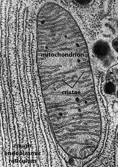
Plant Cell Electron Micrograph Labelled - Candra Kahola Due to the very narrow electron beam, sem micrographs have a large depth of field yielding a characteristic this is an older and noisy micrograph of a common subject for sem micrographs: Figure 7.14 at left a transmission electron micrograph and at right a labeled diagram of a mitochondrion.
Transmission electron microscopy DNA sequencing - Wikipedia Each nucleotide is tagged with a characteristic heavy label, so that they can be distinguished in the transmission electron micrograph. ZS Genetics proposes using three heavy labels: bromine (Z=35), iodine (Z=53), and trichloromethane (total Z=63).
Solved Label the transmission electron micrograph based on - Chegg Expert Answer. nucleus is the house of the genetic material which contains all the h …. View the full answer. Transcribed image text: Label the transmission electron micrograph based on the hints provided Mitochondrion Heterochromatin Plasma cell Nucleus Rough endoplasmic reticulum Nucleolus.
Solved Label the transmission electron micrograph of the - Chegg Question: Label the transmission electron micrograph of the cell. 0 Nucleus rences Mitochondrion Heterochromatin Peroxisome Vesicle ULAR bumit Click and drag each label into the correct category to indicate whether it pertains to the cytoplasm or the plasma membrane. ICF Contacts the ECF Made of proteins and lipids Surrounds the cell Contains lon channels Organelles
To label: MHC-I, MHC-II, pseudopod and a vesicle in the transmission ... Science Biology Microbiology with Diseases by Taxonomy (5th Edition) To label: MHC-I, MHC-II, pseudopod and a vesicle in the transmission electron micrograph of a dendritic cell. Introduction : Dendritic cells are cells of immune system which break down the antigens. Antigen is presented on the surface of the cell along with the major histocompatibiltiy complex.
Animal Cell Electron Microscope Labelled - Q14 Draw a large diagram of ... Animal Cell Electron Microscope Labelled - Q14 Draw a large diagram of an animal cell as seen through ... : The animal cell is more with a transmission electron microscope (tem) and generic contrast staining (osmium, uranyl, lead) of a section through a cell you will not only see.. For example, both animal and plant cells are classified as eukaryotic cells, whereas bacterial cells in a transmission electron microscope, the electron beam penetrates the cell and provides details as you might ...
wgnradio.com › health › uk-early-signs-that-monkeyUK: ‘Early signs’ that monkeypox outbreak may be peaking Aug 05, 2022 · Read More This image provided by the National Institute of Allergy and Infectious Diseases (NIAID) shows a colorized transmission electron micrograph of monkeypox particles (red) found within an infected cell (blue), cultured in the laboratory that was captured and color-enhanced at the NIAID Integrated Research Facility (IRF) in Fort Detrick ...
Electron Micrographs - University of Oklahoma Health Sciences Center Figure 1 Micrograph of a nucleus. 1. Heterochromatin 2. Euchromatin 3. Nucleolus 4. Nucleolar associated chromatin 5. Nuclear envelope Figure 2 Micrograph of a portion of a nucleus: What is the round structure (approximately 3 1/2 inches in diameter) seen in the center of this micrograph? 1. Nucleolar associated chromatin 2.
Label the transmission electron micrograph of the nucleus. - Transtutors Label the transmission electron micrograph of the cell. 0 Nucleus rences Mitochondrion Heterochromatin Peroxisome Vesicle ULAR bumit Click and drag each label into the correct category to indicate whether it pertains to the cytoplasm or the plasma...
Study Chapter 14 & 15 Flashcards Flashcards | Quizlet Key concepts: Physical Barriers Of Innate Immunity Terminal Bronchioles Divide Into Mast Cells And Basophils Terms in this set (101) B-cells may be stimulated to transform into memory cells. True Viruses and self-proteins are examples of proteins produced inside of the cell. True Foreign antigens presented on class I MHC molecules...
Transmission Electron Microscope (TEM)- Definition, Principle, Images Parts of Transmission Electron Microscope (TEM) Their working mechanism is enabled by the high-resolution power they produce which allows it to be used in a wide variety of fields. It has three working parts which include: Electron gun Image producing system Image recording system Electron gun
Label This Transmission Electron Micrograph - Kaiden Brown Interpretation of electron micrographs to identify organelles and deduce the functions of specialized cells. Figures label this transmission electron micrograph ( 16, . No microtubule labeling is evident. Label this transmission electron micrograph of relaxed sarcomeres by clicking and dragging the labels to the correct location .
Electron Microscopic Study of Cell and Organelles - GK SCIENTIST Some of the membranes of endoplasmic reticulum open out in the cell membrane and the others in the nuclear membrane. Golgi apparatus in cytoplasm appears, under an electron microscope, as a pile of 2-layered flattened sacs called cisternae or saccules, lying one within the other.The sacs have flattened or swollen ends near which lie the vesicles of varying size and shape.
#6 Summary of Cell structure | Biology Notes for A level - Blogger 9 The electron micrograph shows part of a secretory cell from the pancreas. The secretory vesicles are Golgi vesicles and appear as dark round structures. The magnification is x 8 000. a Copy and complete the table. Use a ruler to help you find the actual sizes of the structures. Give your answers in micro metres.
Labeled Plant Cell Under Electron Microscope : The Cell Form 1 Biology ... Under a microscope, epithelial cells are readily distinguished by the following features: The electron microscope can achieve a resolution of up to 100 picometers, allowing eukaryotic cells, prokaryotic cells, viruses, ribosomes, and even single atoms to be visualized (note the in order for dna to be clearly visualized under an electron microscope, it must be labeled with heavy atoms.















Post a Comment for "42 label the transmission electron micrograph of the cell."