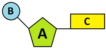
39 label the diagram of the nucleotides below
Topic 7.3, 7.4, 7.5: DNA Replication, Transcription, Translation ... two polymers shown; arranged in a double helix; sugar shown connected to base; sugar-phosphate backbone shown; If only one nucleotide is drawn, award [2 max] sugar identified as deoxyribose; hydrogen bonding between bases shown; diagram shows complementary base pairing / A bonded to T, C with G; Award previous mark if bases (unlabelled) are shown in the diagram but the complementary base ... All About PCR - Beta - University of Utah It can only attach new nucleotides to an existing string of nucleotides. A cell and PCR have different ways of getting started. ... But heating to 95° C, just 5 degrees below boiling, kills most cells. Fortunately, you don’t have to boil DNA to separate its strands. Cells have other ways of doing this, and at much lower temperatures. Your ...
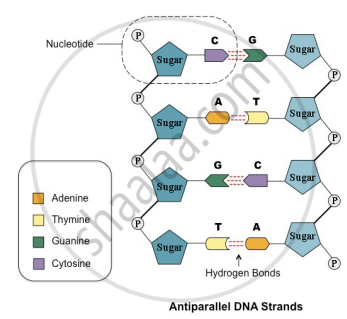
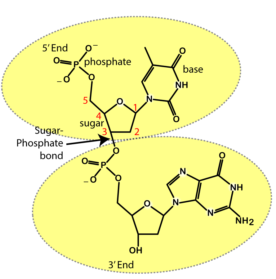
Flow of Genetic Information Kit© Replication Activity Guide Student ... Circle and label the 3' carbon and the 5' carbon in the DNA nucleotide shown in the diagram to the right. ... Examine the detailed diagram of the DNA model below. Double-stranded DNA is composed of two anti-parallel strands! ... Nucleotides are added at an approximate rate of 50 nucleotides per second in eukaryotic cells. The human genome ...
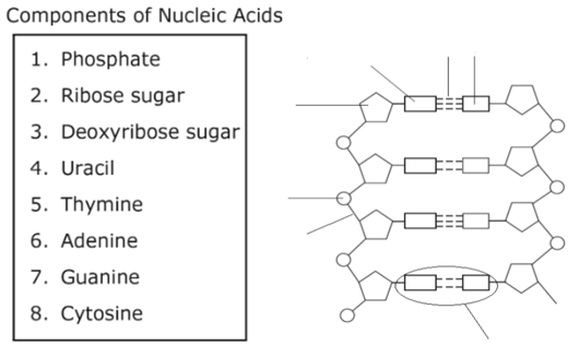
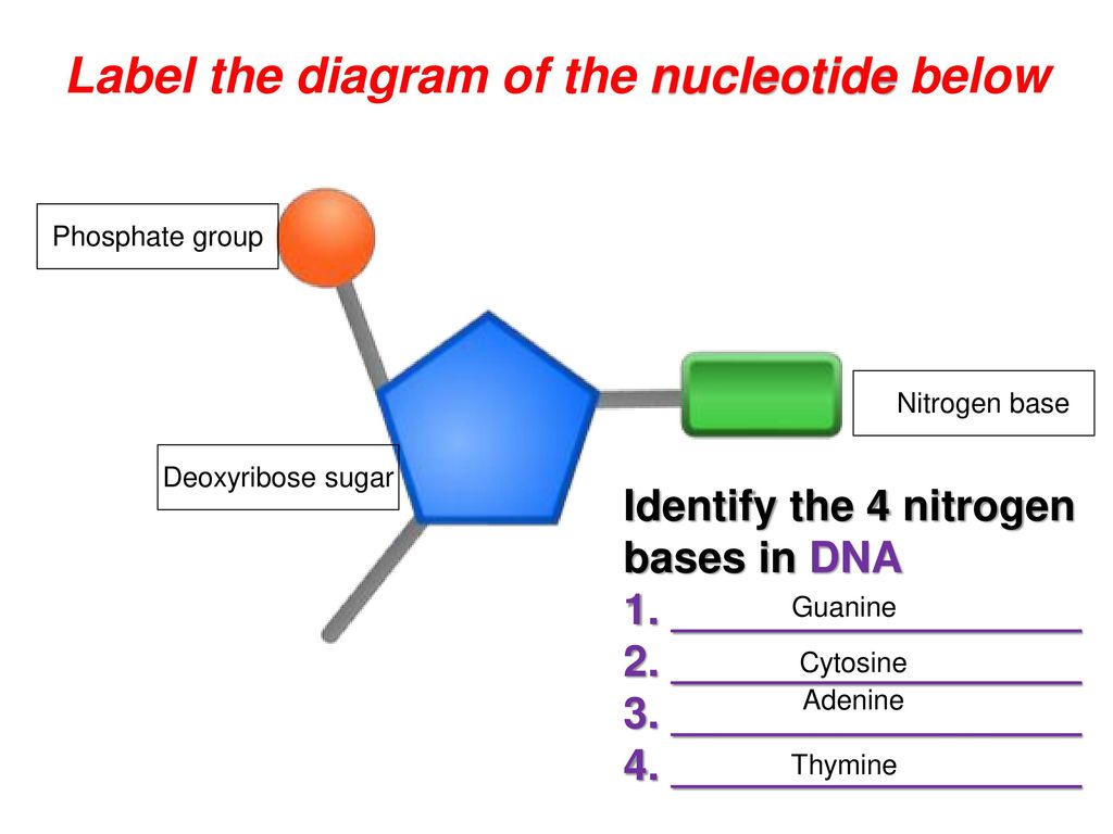
Label the diagram of the nucleotides below
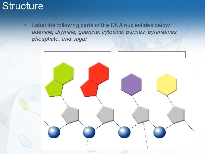
Sketch And Label A Nucleotide : Solved Problem 1 What Are The ... - Blogger The general structural representation of a nucleotide is shown below. A bond between nucleotides in rna and dna molecules. Draw a nucleotide and label the three main parts. See below the above structure is a color (magenta)nucleotide. Using arrows and brackets, identify and label at least one example of each of the following. DNA: Structure, Forms and Functions (With Diagram) - Biology Discussion These forms are described below: (i) A-DNA or Right-Handed DNA: The A-structure is induced in DNA in 70% to 75% ethanol and is found in fibres of DNA in a dehydrated state. The primary difference between A and B helices lies in the sugar ring conformation (pucker). The sugars are C' 3-endo in the A-form but C' 2 endo in the B-form (Fig. 10.17). Label the parts of the DNA in the diagram given below. Explain the ... DNA is the hereditary material as it contains genetic information. DNA is a large molecule, consisting of millions of nucleotides. Each nucleotide consists of three compounds. Nucleotides in a DNA (a) A sugar molecule - Deoxy Ribose sugar (b) A nitrogenous base [Purines and Pyrimidines] Purines (Adenine and Guanine) Pyrimidines (Cytosine and ...
Label the diagram of the nucleotides below. Nucleotide Structure: DNA Diagram | Science Trends As mentioned, nucleotides have three component parts: a five-sided carbon sugar, a nitrogen-containing base, and a phosphate group. The sugar and phosphate group together to create the sugar phosphate backbone. This is skeleton or foundation of the DNA double helix. 16identify and label the diagram below 17 list the - Course Hero 16.Identify and label the diagram below: 1. Primase joins RNA nucleotides into a primer 2. DNA pol 3 addsDNA nucleotides to the primer, forming Okazakifragment 1. 3. After reaching the next RNA primer to the right, DNApol 3 detaches. 4. After fragment 2 is primed, DNA pol 3 adds DNAnucleotides until it reaches the fragment 1 primer and detaches. 5. DNA Structure - YouTube Learn about the structure of DNA and how to recognize all the parts in this video! Matrix-assisted laser desorption/ionization - Wikipedia In mass spectrometry, matrix-assisted laser desorption/ionization (MALDI) is an ionization technique that uses a laser energy absorbing matrix to create ions from large molecules with minimal fragmentation. It has been applied to the analysis of biomolecules (biopolymers such as DNA, proteins, peptides and carbohydrates) and various organic molecules (such as polymers, …
Microbiology Chapters 9 Flashcards | Quizlet A mouse containing such a transgene will express a hybrid protein X-.GFP only in those tells in which gene X is normally expressed. a. The gene X - GFP fusion gene described could be generated by knocking in GFP coding sequences instead of by random insertion of a transgene. Diagram the knockin construct you could use for this purpose. b. How do you draw a nucleotide and label its three basic parts? Explanation: The above structure is a nucleotide. It consists of a: phosphate group 5-carbon sugar, and nitrogenous base. Answer link Helicase - Wikipedia With the use of specialized mathematical equations, some of these assays can be utilized to determine how many base paired nucleotides a helicase can break per hydrolysis of 1 ATP molecule. Commercially available diagnostic kits are also available. One such kit is the "Trupoint" diagnostic assay from PerkinElmer, Inc. This assay is a time ... PDF Bio 102 Practice Problems Chromosomes and DNA Replication 6. The diagram below shows a DNA molecule in the process of replication. Arrows show the direction of new DNA synthesis. a. Some of the enzymes and features are labeled, but some labels are incomplete or have been omitted. Fill in the boxes with the appropriate labels. b.
Week 1: Protein Synthesis Flashcards | Quizlet Drag the correct labels onto the diagram to identify the structures and molecules involved in translation. The diagram below shows the arrangement of the translation components during initiation. Label each component with the most appropriate and specific label provided. A site: aminoacyl P site: peptidyl E site: exit Biopython - Quick Guide - tutorialspoint.com The process of creating a diagram generally follows the below simple pattern − Create a FeatureSet for each separate set of features you want to display, and add Bio.SeqFeature objects to them. Create a GraphSet for each graph you want to display, and add graph data to them. Use the components in the list below to label the diagram of a ... Use the components in the list below to label the diagram of a replication fork in the figure. a.) DNA polymerase b.) single-stranded binding protein c.) Okazaki fragment d.) primase e.) sliding clamp f.) RNA primer g.) DNA helicase 138. Telomeres serve as caps at the ends of linear chromosomes. iCn3D: Web-based 3D Structure Viewer - National Center for ... What is iCn3D Structure Viewer? "I see in 3D" (iCn3D) Structure Viewer is not only a web-based 3D viewer, but also a structure analysis tool interactively or in the batch mode using NodeJS scripts based on the npm package icn3d. iCn3D synchronizes the display of 3D structure, 2D interaction, and 1D sequences and annotations.
6A DNA Structure Review | Biology Quiz - Quizizz
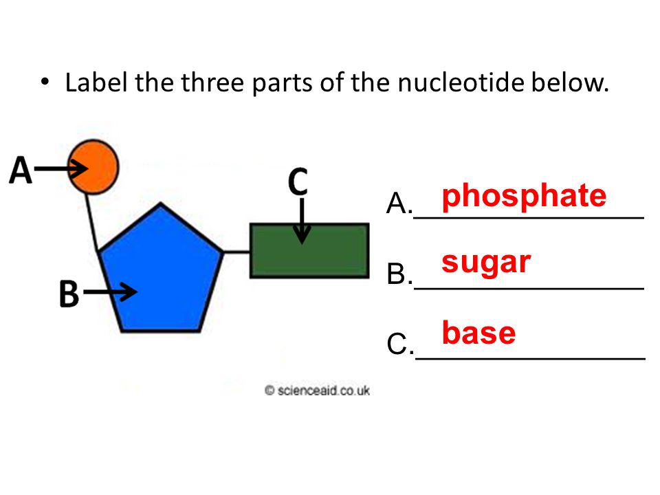
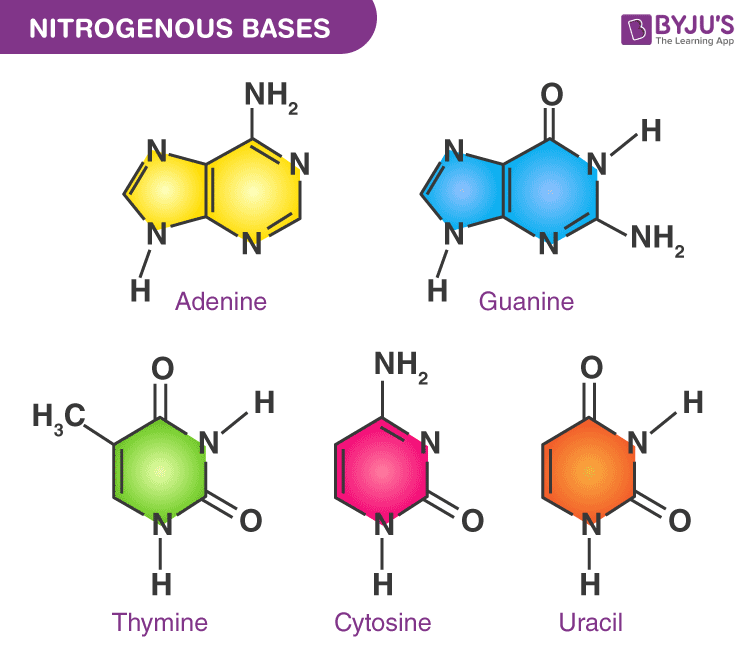
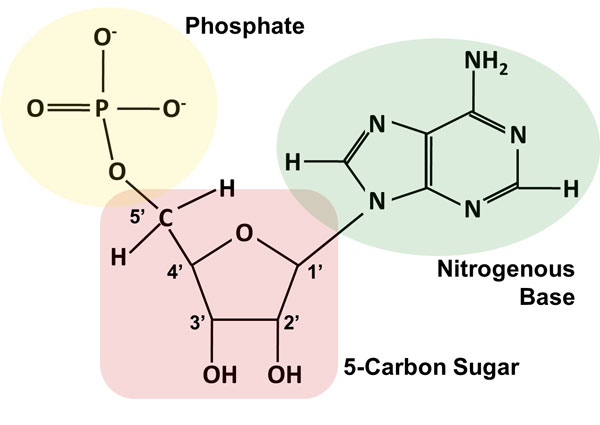
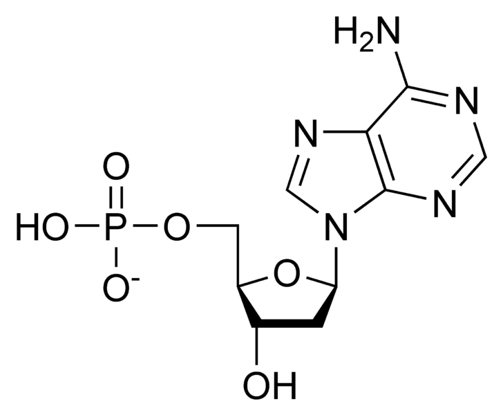
Nucleotide - Definition, Structure (3 Parts), Examples & Function A nucleotide is made up of three parts: a phosphate group, a 5-carbon sugar, and a nitrogenous base. The four nitrogenous bases in DNA are adenine, cytosine, guanine, and thymine. RNA contains uracil, instead of thymine. A nucleotide within a chain makes up the genetic material of all known living things.
Interim 2 review. - ppt video online download
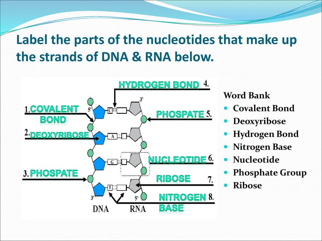
Nucleotide - Genome.gov A nucleotide is the basic building block of nucleic acids (RNA and DNA). A nucleotide consists of a sugar molecule (either ribose in RNA or deoxyribose in DNA) attached to a phosphate group and a nitrogen-containing base. The bases used in DNA are adenine (A), cytosine (C), guanine (G) and thymine (T). In RNA, the base uracil (U) takes the ...

How Well You Know DNA Structure? Quiz - ProProfs Quiz
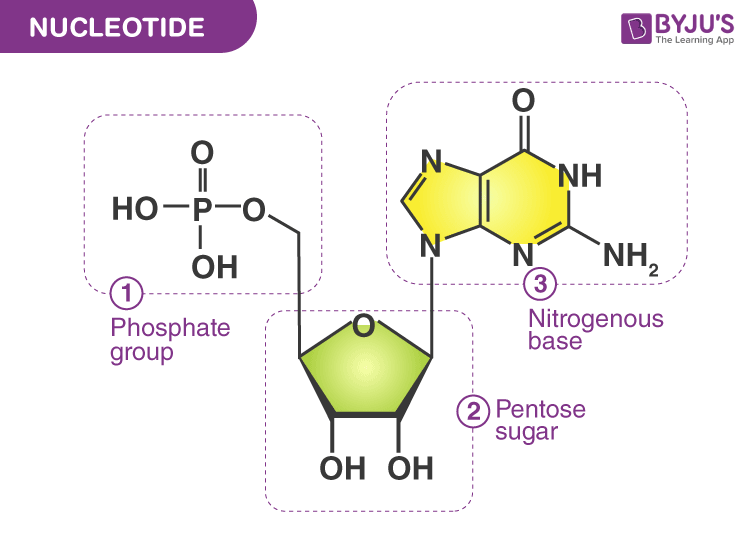
Nucleotide: Structure, Examples and Function - BYJUS Sugar: A nucleotide contains a pentose sugar. DNA (Deoxyribonucleic acid) contains deoxyribose sugar and RNA (Ribonucleic acid) contains a ribose sugar. A Nitrogenous base attached with the sugar is called "Nucleoside". 3. Phosphate: Phosphate is attached to the sugar of nucleoside by an ester bond with the 5 th C hydroxyl group.

9.1 The Structure of DNA – Concepts of Biology – 1st Canadian ...
GRADE 12 LIFE SCIENCES LEARNER NOTES - Mail & Guardian 3.1 Label the molecules indicated by 2 and 3. (2) 3.2 Using the letters of the genetic code, write down the complementary nitrogenous bases on strand 1 of the DNA double helix, starting from the top. (3) (Remember: A=T/U and G=C) 3.3 Use the table below to determine which three amino acids in the diagram are represented by 4, 5 and 6. (3 x 2) (6)

2.6 and 2.7 Paper 2 Questions Flashcards | Quizlet
Draw a well labelled diagram of an eukaryotic nucleus. How is it ... The diagram given below represents a plant cell after being placed in a strong sugar solution. Study the diagram and answer the questions that follow: ... Draw a diagram of typical cell and label the following parts in it. Cell membrane Vacuole Nucleus Endoplasmic reticulum Mitochondria Golgi body. Medium. View solution > View more.
DNA Structure and Function DNA Structure and Function
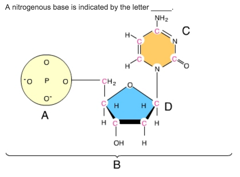
Solved Directions: Label the diagram below with the | Chegg.com Directions: Label the diagram below with the following choices: • Nucleotide • Deoxyribose • Phosphate group • Base pair • Hydrogen bond • Nitrogenous base 7. A nucleotide is made of three parts: a _________________________ group, a five carbon _________________________, and a _________________________ base. 8.

Nucleotides and Nucleic acids Flashcards | Quizlet
DNA Molecule Label Diagram | Quizlet Molecule found on the side of a DNA molecule Double Helix two strands of nucleotides wound about each other; structure of DNA Thymine the nucleotide that hydrogen bonds with the nucleotide adenine in DNA Adenine the nucleotide that hydrogen bonds with the nucleotide thymine in DNA or with uracil in RNA Guamine
DNA, RNA & Protein Synthesis - ppt download
What are the Three Parts of a Nucleotide? | Albert.io Nucleotides are made up of 3 parts. The first is a distinct nitrogenous base, which is adenine, cytosine, guanine or thymine. In RNA, thymine is replaced by uracil. These nitrogenous bases are either purines or pyrimidines. Base pairs are formed when adenine forms a hydrogen bond with thymine, or cytosine forms a hydrogen bond with guanine.
Label the parts of the DNA in the diagram given below ...
Top 8 Types of Genetic Markers (With Applications) When tested with a group of independent VNTR markers, the likelihood of two unrelated individuals having the same allelic pattern is extremely improbable. In the example considered in the diagram below locus A is a tandem repeat of the motif GC: there are four alleles, with two, three, four, or five repeats (A2, A3, A4, and A5, respectively).
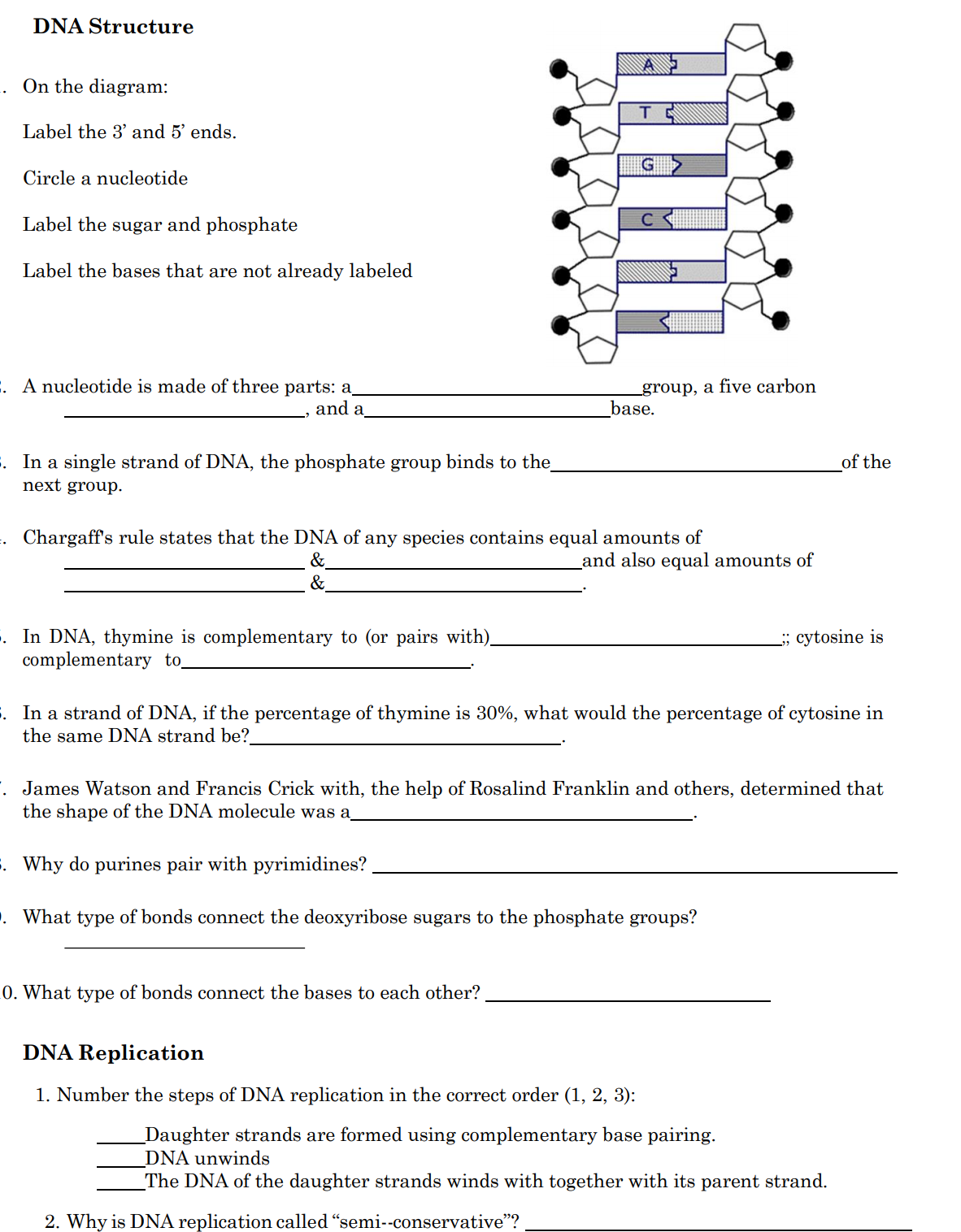
Answered: DNA Structure On the diagram: Label the… | bartleby
Draw And Label A Single Dna Nucleotide / Draw And Label Schematic ... The general structural representation of a nucleotide is shown below. Draw a nucleotide and label the three main parts. The phosphodiester bonds that join one dna nucleotide to another always link . Nucleotides are the building blocks of the dna and rna used as genetic material. The dna molecule actually consists of .

Label the parts of the drawing below. include all of these ...
(Solved) - Directions: Label the diagram below with the following ... A nucleotide is made of three ...
Chapter 8 From DNA to Proteins - ppt video online download
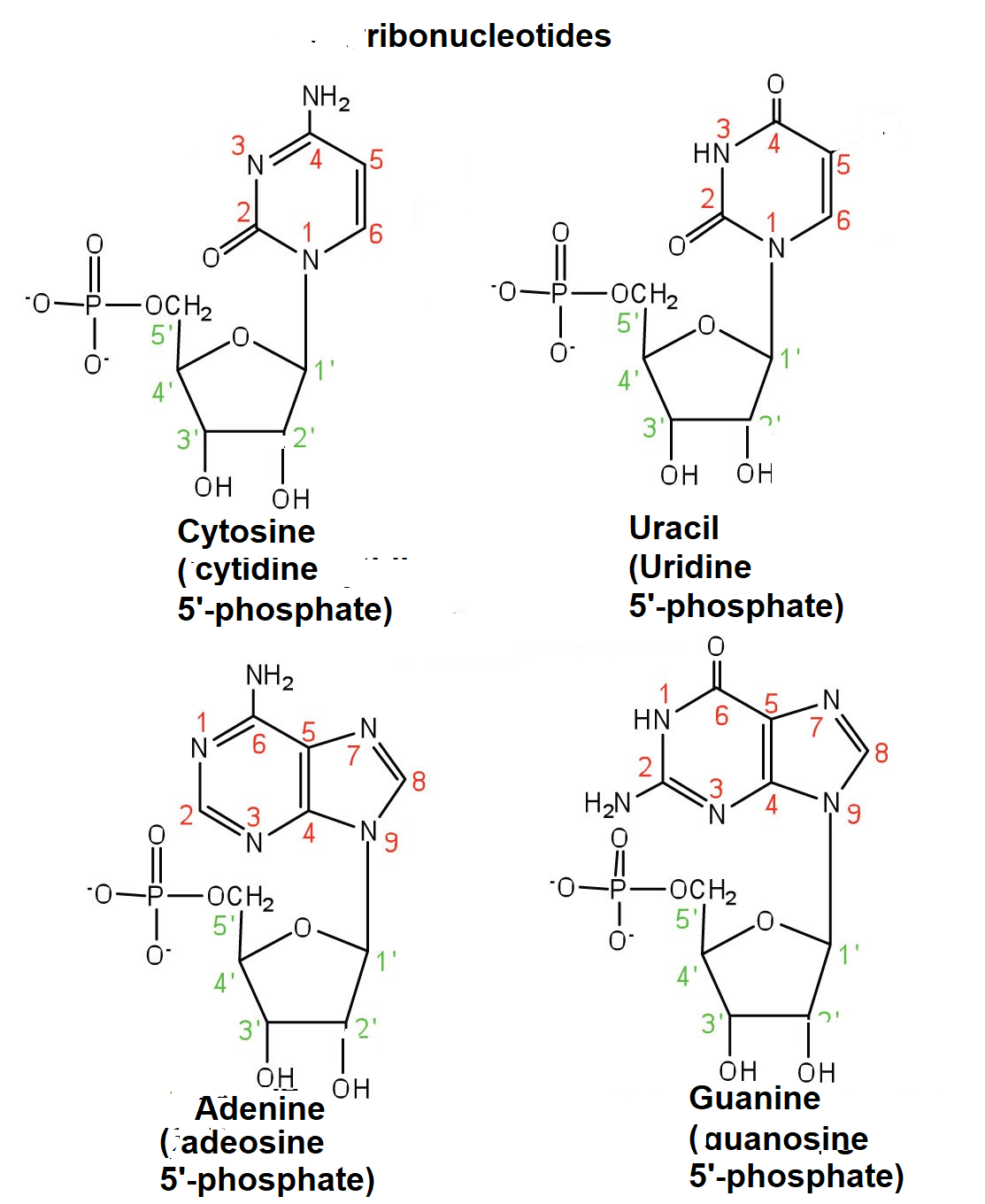
3 Parts of a Nucleotide and How They Are Connected - ThoughtCo Cytosine, thymine, and uracil are pyrimidines. In DNA, the bases are adenine (A), thymine (T), guanine (G), and cytosine (C). In RNA, the bases are adenine, guanine, uracil, and cytosine. Pentose Sugar In DNA, the sugar is 2'-deoxyribose. In RNA, the sugar is ribose. Both ribose and deoxyribose are 5-carbon sugars.
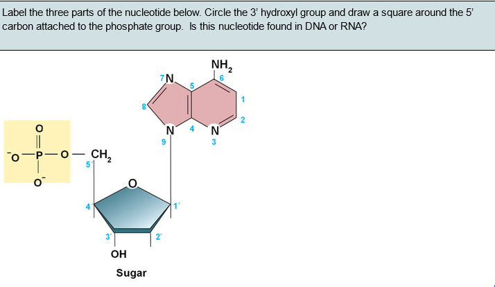
Solved Label the three parts of the nucleotide below. Circle ...
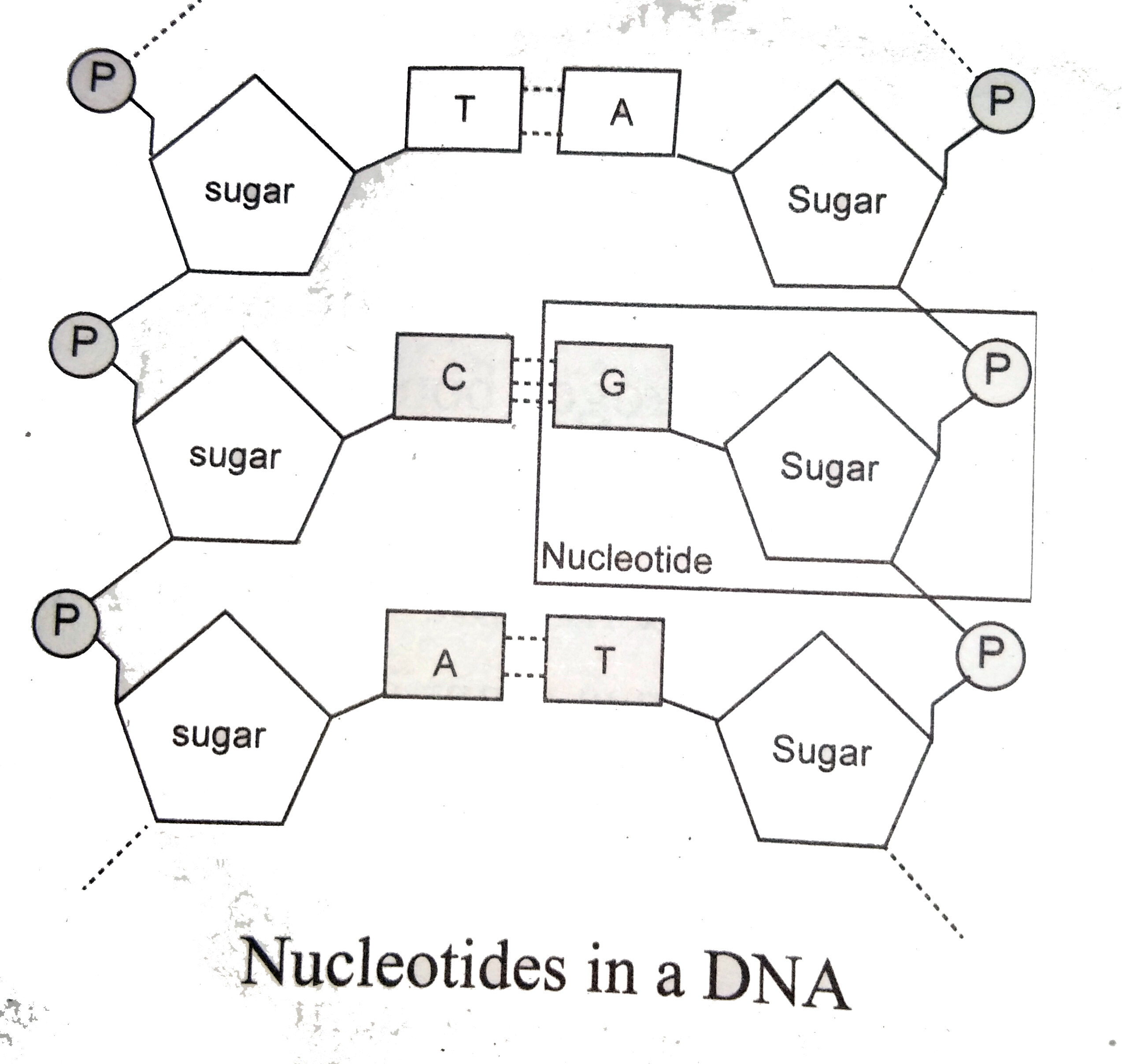
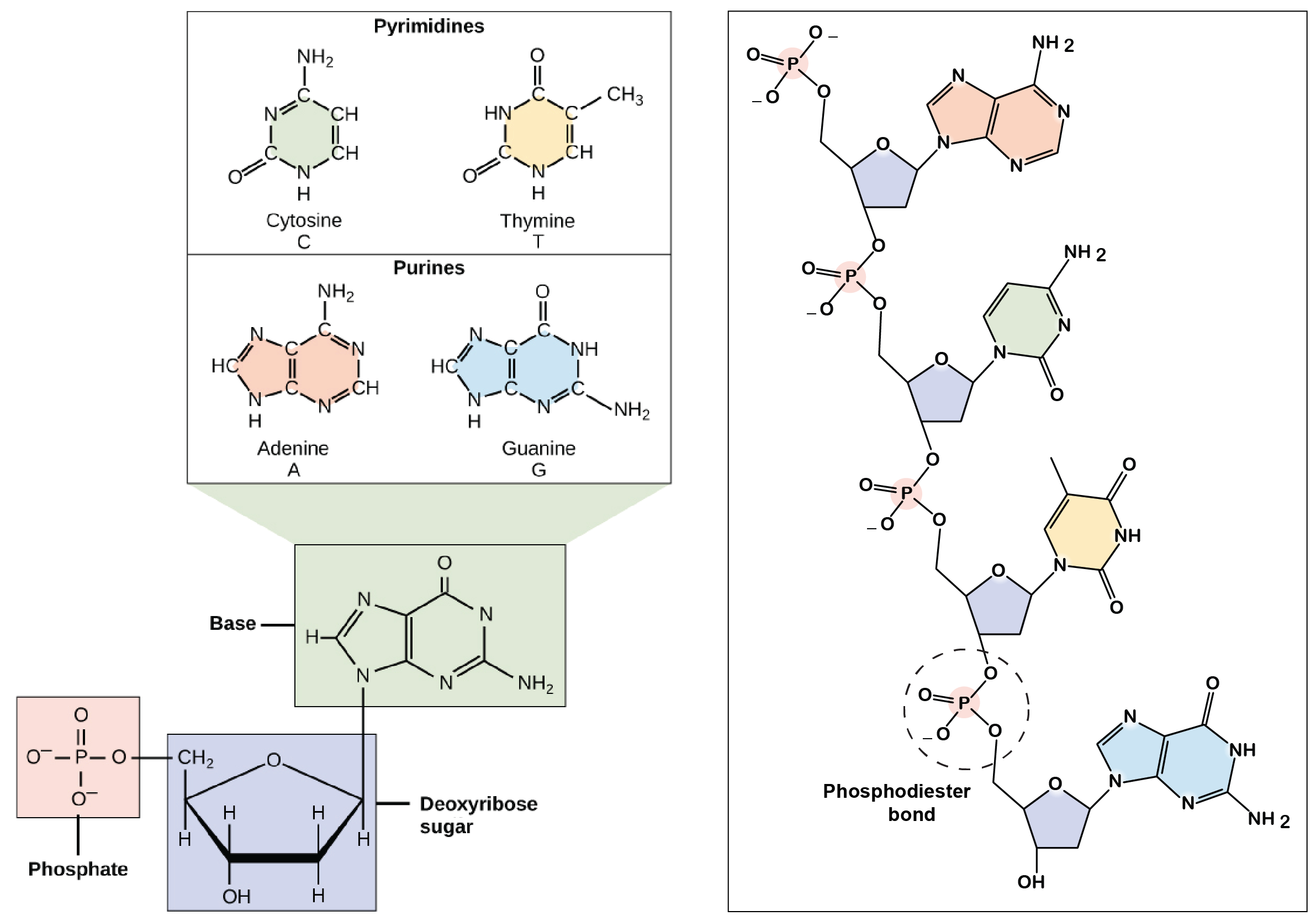
The Structure of DNA - University of Arizona The boxed area at the lower left encloses one nucleotide. Each nucleotide is itself make of three subunits: A five carbon sugar called deoxyribose (Labeled S) A phosphate group (a phosphorous atom surrounded by four oxygen atoms.) (Labeled P) And one of four nitrogen-containing molecules called nucleotides . (Labeled A, T, C, or G)

How do you draw a nucleotide and label its three basic parts ...
Solved ta Directions: Label the diagram below with the - Chegg Transcribed image text: ta Directions: Label the diagram below with the following choices: • Nucleotide • Deoxyribose • Phosphate group • Base pair • Hydrogen bond • Nitrogenous base ca sh fn DNA Molecule: Two Views 2. H H-C H- HE HC -H- -H SHO 5. THO 3. Directions: Complete each sentence. 7. Guanine, cytosine, thymine, and are the four in DNA. 8.
9.1 The Structure of DNA – Concepts of Biology – 1st Canadian ...
FortéBio Bio-layer Interferometry Kinetic Analysis Tutorial Nucleotides (DNA) Small Molecules Atoms 200 nm 1000 nm 0.1 nm 1 1,000 100,000 75 nm 1,000,000 ... Right click on highlighted wells or select from options below plate to select well type 2 1. 33 ... biosensors in every other column Follow the diagram to load your biosensors Biosensor Location #1 will be used in this mode with re-racking 8 ...
Discovery of the structure of DNA (article) | Khan Academy
(Solved) - Directions: Label the diagram below with the following ... Directions: Label the diagram ...

Nucleic acids
DNA and RNA (With Diagram) - Biology Discussion These constitute 5-10% of the total RNA is the cell. m-RNA is synthesized in nucleolus and after taking genetic information from DNA goes into the cytoplasm and helps in the formation, of specific protein. m-RNA are short lived. Sequence of 3 bases of nucleotides in m-RNA molecule constitutes a codon.
Exam 3: Chs. 5 (DNA Structure and Replication Machinery) & 16 ...
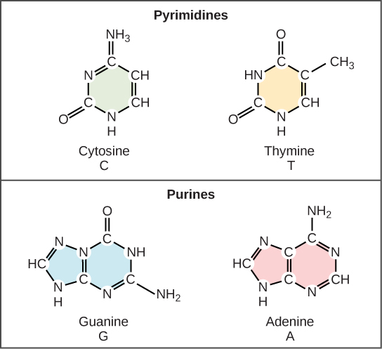
9.1 The Structure of DNA - Concepts of Biology - 1st Canadian Edition The DNA molecule is a polymer of nucleotides. Each nucleotide is composed of a nitrogenous base, a five-carbon sugar (deoxyribose), and a phosphate group. There are four nitrogenous bases in DNA, two purines (adenine and guanine) and two pyrimidines (cytosine and thymine). A DNA molecule is composed of two strands.
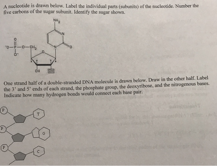
Solved A nucleotide is drawn below. Label the individual ...
Labeling Oligonucleotides and Nucleic Acids—Section 8.2 The ARES DNA Labeling Kits use a two-step method to label DNA. Step 1) The aminoallyl dUTP is enzymatically incorporated. Step 2) A reactive fluorophore is used to label the incorporated aminoallyl group. Figure 8.2.9 Change in signal brightness with level of labeling for Alexa Fluor versus Cy dye-labeled DNA.

Nucleotides | BioNinja
Label the parts of the DNA in the diagram given below. Explain the ... DNA is the hereditary material as it contains genetic information. DNA is a large molecule, consisting of millions of nucleotides. Each nucleotide consists of three compounds. Nucleotides in a DNA (a) A sugar molecule - Deoxy Ribose sugar (b) A nitrogenous base [Purines and Pyrimidines] Purines (Adenine and Guanine) Pyrimidines (Cytosine and ...
DNA Nucleotide Labeled and Unlabeled by Ms Mares Store | TpT
DNA: Structure, Forms and Functions (With Diagram) - Biology Discussion These forms are described below: (i) A-DNA or Right-Handed DNA: The A-structure is induced in DNA in 70% to 75% ethanol and is found in fibres of DNA in a dehydrated state. The primary difference between A and B helices lies in the sugar ring conformation (pucker). The sugars are C' 3-endo in the A-form but C' 2 endo in the B-form (Fig. 10.17).
Label the parts of the DNA in the diagram given below ...
Sketch And Label A Nucleotide : Solved Problem 1 What Are The ... - Blogger The general structural representation of a nucleotide is shown below. A bond between nucleotides in rna and dna molecules. Draw a nucleotide and label the three main parts. See below the above structure is a color (magenta)nucleotide. Using arrows and brackets, identify and label at least one example of each of the following.
Nucleotide: Structure, Examples and Function
8.2 Reading worksheet

Nucleic Acids Flashcards | Quizlet

Draw a schematic representation of dinucleotide Label the ...
How do you draw a nucleotide and label its three basic parts ...
28.1: Nucleotides and Nucleic Acids - Chemistry LibreTexts

Label the parts of the DNA in the diagram given below ...
Nucleotide: Structure, Examples and Function

DNA comprehensive.doc - Name_Zacery Coleman_ Date:_ Mod:_ ...

Solved Correctly label the parts of the two-nucleotide ...
DNA Structure (Interactive Tutorial) – learn-biology

Selina Solutions Concise Biology Class 10 Chapter 2 Structure ...

Nucleic Acids are composed of nucleotides. The DNA and RNA ...
DNA - structure
What are the Three Parts of a Nucleotide? | Albert.io
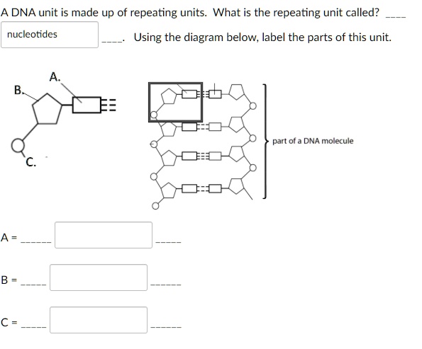
SOLVED:A DNA unit is made up of repeating units What is the ...

Given below is a schematic diagram of a portion of DNA.a) How ...
Post a Comment for "39 label the diagram of the nucleotides below"