44 label the photomicrograph of the sebaceous gland
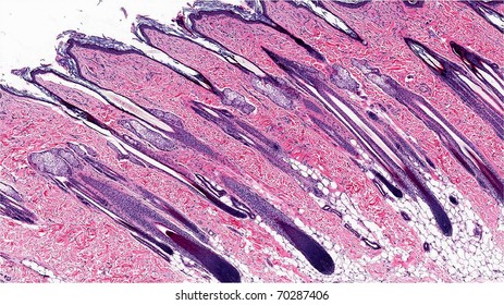
Integumentary System HW_answers.docx - Course Hero Hair follicle , sebaceous gland , sweat gland. 11. cells indicated. 12. In the micrograph of a longitudinal section through a nail, identify the parts of a nail and surrounding structures. 13. Label the structures of the fingernail in a lateral view. 14. ... Label the photomicrograph of the sebaceous gland. 20. unit 4 lab.docx - LAB Unit 4 EXERCISE 7: The ... - Course Hero FIGURE 7.4: Diagram of the skin and accessory structures. • apocrine (AP-oh-krin) sweat gland • arrector pili (PIE-lee) muscle • eccrine (EK-rin) sweat gland • hair bulb • hair follicle • hair root • hair shaft • papilla (puh-PILL-uh) of hair • sebaceous (se-BAY-shus) gland 1. Hair shaft 2. Hair root 3. Sebaceous glands 4. Arrector pili muscle 5.
photomicrograph of a sectioned sebaceous gland - Quizlet Start studying photomicrograph of a sectioned sebaceous gland. Learn vocabulary, terms, and more with flashcards, games, and other study tools.

Label the photomicrograph of the sebaceous gland
Label The Photomicrograph Of Thin Skin Quizlet - Skin Labeling Review ... Sebaceous Oil Gland Hair Follicle Ppt Download from slideplayer.com Label the photomicrograph of thick skin. In the photomicrograph of a portion of thick skin shown below, . 28) in the diagram of skin shown below, which labeled structure generates fingerprints? Learn vocabulary, terms, and more with flashcards, games, and other study tools. (Get Answer) - Label The Photomicrograph Of The Skin And Its Accessory ... Activity 4 Differentiating Sebaceous and Sweat Glands Microscopically Using the slide thin skin with hairs and the photomicrographs of cutaneous glands (Figure 7.6) as a guide, identify sebaceous and eccrine sweat glands. What characteristics... Photomicrograph Of Human Scalp Showing Epidermis Dermis ... - Getty Images View top-quality stock photos of Photomicrograph Of Human Scalp Showing Epidermis Dermis Subcutaneous Layer Hair Shaft Hair Follicles Adipose Tissue Blood Vessels And Sebaceous Glands 10x. Find premium, high-resolution stock photography at Getty Images.
Label the photomicrograph of the sebaceous gland. Figure 7.4 Photomicrograph of the skin and accessory ... - Quizlet Start studying Figure 7.4 Photomicrograph of the skin and accessory structures. Learn vocabulary, terms, and more with flashcards, games, and other study tools. ... Sebaceous Gland. Oil glands that surround hair follicles; secrete oils that lubricates skin, hair, and into the neck of the hair follicle. ... Final Exam A&P 1 Flashcards | Quizlet Label the structures of the skin and subcutaneous tissue. Label the photomicrograph of thick skin. Epidermis, stratum corneum, stratum lucidum, stratum granulosum, stratum spinosum, stratum basale, dermis Label the photomicrograph of thin skin Hair shaft, epidermis, dermal root sheath, sebaceous gland, dermis, hair matrix Anatomy and Physiology Homework Chapter 6 Flashcards - Quizlet Sebaceous glands produce an oily secretion called sebum. They are flask-shaped, with short ducts that usually open into a hair follicle, although some of them open directly onto the skin surface. ... Label the photomicrograph of the sebaceous gland.-Duct of sebaceous gland-Secretory cell-Sebaceous gland-Basal cell-Hair follicle-Hair follicle ... skin labeling quizlet - gooddog.co.za The following is an overview of the male reproductive anatomy: Scrotum. Your Skills & Rank. below according to the structure/area they describe. Label the structures of the skin and. Label the photomicrograph of thin skin. You need to get 100% to score the 20 points available.
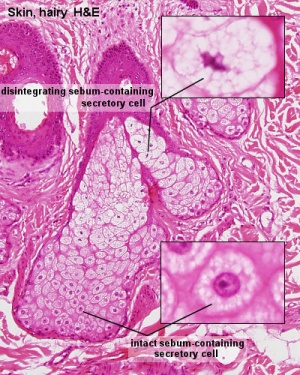
PDF Name the Condition - Dr. Scott Croes' Website the cartoon and the photomicrograph. Name the 4 layers of thin skin in both the cartoon and the photomicrograph. •Name the Layers of skin and label the dermal papilla and dermis •Name the Layers of skin and label the dermal papilla ... Sebaceous gland • Identify the following: Dermal papilla; Melanocyte; Label The Photomicrograph Based On The Hints Provided / Endocrine Lab ... Label the photomicrograph based on the hints provided zona fasciculata suprarenal gland zona reticularis capillary medulla dr thomas . Spleen capsule capsule white pulp. Name and describe the different layers of the adrenal . Medulla capillary zona fasciculara suprarenal gland zona reticularis. Label The Photomicrograph Of The Sebaceous Gland ... Apr 01, 2022 · Label the photomicrograph of the sebaceous gland. These structures embryologically originate from . If the gland become blocked, the sebum can be forced out into the dermis, where it elicits an inflammatory response. Label the photomicrograph of the skin and its accessory structures. Epidermis hair follicle duct of sebaceous gland sebaceous gland. Chapter 6 - Labeling Parts of Skin - sweat gland blood... sweat gland blood vessels sweat sweat gland pore muscle layer Labeling Parts of the Skin Identify the layers of skin. dermis stratum basale stratum spinosum stratum lucidum stratum corneum stratum granulosum basement membrane Identify the parts of this photomicrograph of skin: sebaceous gland hair shaft hair follicle dermis epidermis
Mandala Pferde Zum Ausmalen / Mandala Ausmalbilder Entspannung ... Hair sebaceous gland dermis hair follicle epidermis duct of sebaceous. Label the photomicrograph... Crayola Crayon Wrapper Template : Free Printable Crayola Crayon Templates Crayon Template Templates Printable Free Crayola Crayons. Crayola® crayon labels cannot be personalized. Add your name to your crayons or make them kawaii cute with these ... (Get Answer) - Label the photomicrograph in Figure 7.4. Examine a slide ... Label the layers of the skin on the diagram and the photograph. Be able to identify the layers on a microscope slide. Look at the skin slide under a microscope. a) Epidermis 1) Stratum corneum ii) Stratum lucidum 111) Stratum granulosum iv) Stratum... Posted 11 months ago Recent Questions in Basics of Statistics Q: Solved Label the photomicrograph of the sebaceous gland ... Experts are tested by Chegg as specialists in their subject area. We review their content and use your feedback to keep the quality high. 100% (28 ratings) Transcribed image text: Label the photomicrograph of the sebaceous gland. Previous question Next question. Answered: • hair bulbs • hair follicle • hair… | bartleby Solution for • hair bulbs • hair follicle • hair root • papilla of hair • sebaceous gland 1 1 2 2 3 3 4 5 4 5 10x Courtesy Michael Ross, University of Florida
Skin - Hair Follicles and Oil Glands - Histology | Histology - Skin ...
Bio Lab Chapter 6 Quiz Flashcards | Quizlet -hair follicle and sebaceous gland Identify the type of tissue that composes the epidermis of the skin. stratified squamous epithelial tissue Identify the structures of the dermis. dense connective tissue with fibers oriented in many directions dense irregular loose connective tissue characterized by long, thin dark fiber areolar tissue
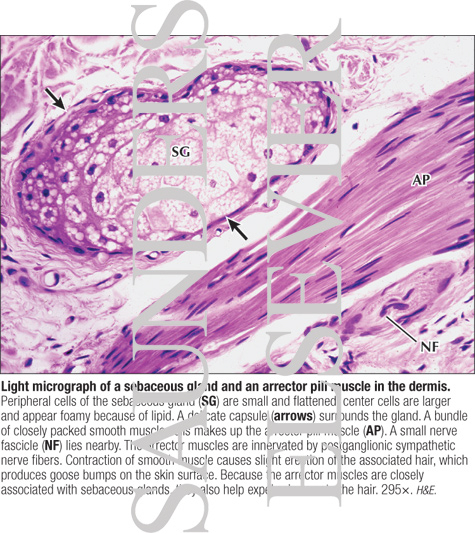
Light Micrograph of a Sebaceous Gland and an Arrector Pili Muscle In ...
CH 5 Integument - CHAPTER 5 INTEGUMENT Skin(Integument ... - Course Hero Figure 5.2a The main structural features of the skin epidermis. Dermis Stratum spinosum Several layers of keratinocytes unified by desmosomes. Cells contain thick bundles of intermediate filaments made of pre-keratin. Stratum basale Deepest epidermal layer; one row of actively mitotic stem cells; some newly formed cells become part of the more superficial layers.

Skin Anatomy and Wound Healing - Dermatology - Medbullets Step 2/3
HelenakruwAllison Label the Photomicrograph of the Sebaceous Gland. By fu_Margaret102 10 May, 2022 Post a Comment ... Label the Photomicrograph of the Sebaceous Gland. These patients often complain of. Below the dermis is anoth… Ucapan Selamat Hari Raya Aidilfitri in English
Integumentary System - Gland Development - Embryology
sudoriferous glands location - hairstylezz.com Sudoriferous glands are also called sweat glands. Sebaceous Glands. duct gland; exocrine; exocrine gland (a gland that secretes externally through a duct) Hyponyms (each of the following is a kind of "sudoriferous gland"): apocrine gland (a large sweat gland that produces both a fluid and an apocrine secretion; in human â ¦ When internal ...
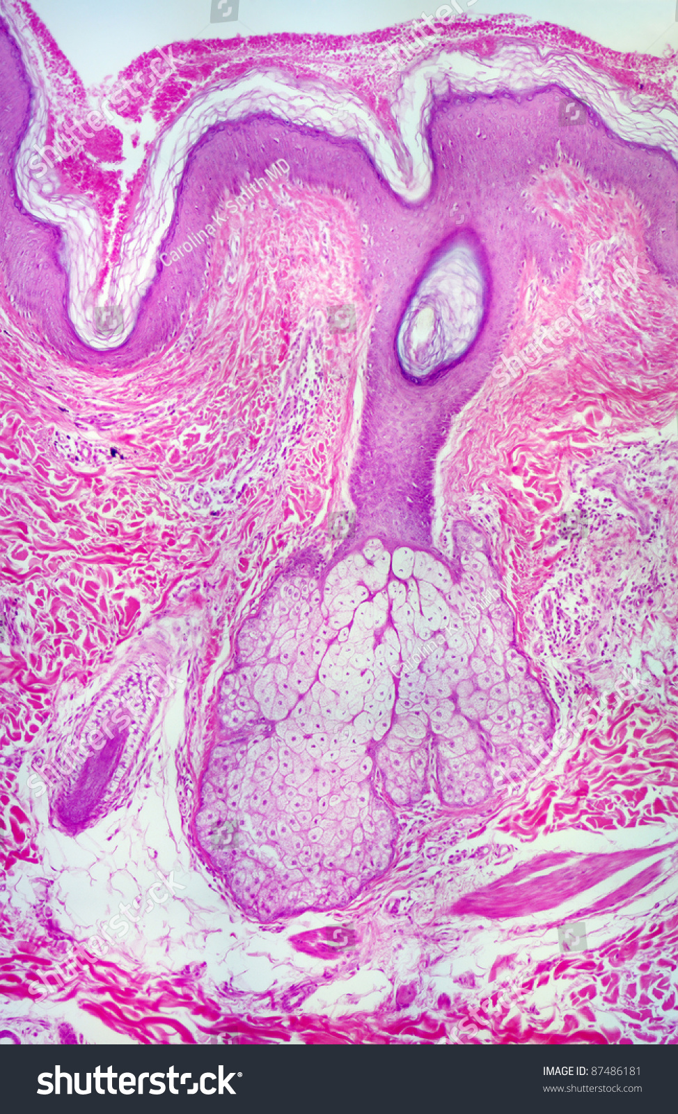
Real Human Sebaceous Sweat Gland Photomicrograph Stock Photo 87486181 ...
Solved Label the photomicrograph of the skin and its | Chegg.com Label the photomicrograph of the skin and its accessory structures Epidermis Duct of sebaceous gland Hair follicle Sebaceous gland Show transcribed image text Expert Answer 2. The picture here demonstrates the pseudostratified columnar epithelium.

Human Scalp Shows Hair Follicle Hair Shaft Sebaceous Glands Dermis ...
Solved > Question 31 points Label the photomicrograph of ... - ScholarOn Question 31 points Label the photomicrograph of thin skin. Hair Follicle . Not my Question Bookmark. Flag Content. ... 31 points Label the photomicrograph of thin skin. Hair Follicle Hair Dermis Sebaceous gland Duct of sebaceous gland Reset zoom. Solution. 5 (1 Ratings ) Solved. Biology 2 Years Ago 68 Views. This Question has Been Answered ...

c-MYC-Induced Sebaceous Gland Differentiation Is Controlled by an ...
Sebaceous Gland Label The Photomicrograph Of Thin Skin / Accessory ... Sebaceous Gland Label The Photomicrograph Of Thin Skin / Accessory Structures Of The Skin Anatomy And Physiology. This is the spellchex dictionary for online spell checking. Endocrine mucin producing sweat gland carcinomas always categorical a minimum of one.
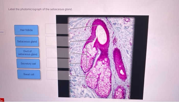
29 Label The Photomicrograph Of The Sebaceous Gland. - Modern Labels ...
Photomicrograph Of Human Scalp Showing Epidermis Dermis ... - Getty Images View top-quality stock photos of Photomicrograph Of Human Scalp Showing Epidermis Dermis Subcutaneous Layer Hair Shaft Hair Follicles Adipose Tissue Blood Vessels And Sebaceous Glands 10x. Find premium, high-resolution stock photography at Getty Images.
30 Label The Photomicrograph Of The Sebaceous Gland. - Labels Design ...
(Get Answer) - Label The Photomicrograph Of The Skin And Its Accessory ... Activity 4 Differentiating Sebaceous and Sweat Glands Microscopically Using the slide thin skin with hairs and the photomicrographs of cutaneous glands (Figure 7.6) as a guide, identify sebaceous and eccrine sweat glands. What characteristics...
Post a Comment for "44 label the photomicrograph of the sebaceous gland"